Home » Without Label » Compact Bone Diagram / Schematic Diagram of Compact and Spongy Bones. Schematic ... - (b) in this micrograph of the osteon, you can clearly see the concentric lamellae and central canals.
Compact Bone Diagram / Schematic Diagram of Compact and Spongy Bones. Schematic ... - (b) in this micrograph of the osteon, you can clearly see the concentric lamellae and central canals.
Compact Bone Diagram / Schematic Diagram of Compact and Spongy Bones. Schematic ... - (b) in this micrograph of the osteon, you can clearly see the concentric lamellae and central canals.. The remainder of the bone is formed by cancellous or spongy bone. Andrew kirmayer a diagram of the anatomy of a bone, showing the compact bone. Compact bone is hard and forms the outer layer of any bone. Bones are hard organs inside our bodies that make up our skeletal system. The key difference between compact bone and spongy bone is that the compact bone is a tough and heavy bone that forms the diaphysis of long bones while the spongy bone is a soft and light bone that forms the epiphysis of long bones.
The latter helps save materials, and provide movement to. Compact bone is formed from a number of osteons, which are circular units of bone material and blood vessels. The spongy bone crowds nearby blood vessels, which eventually condense into red bone marrow ( figure 6.4.1 d ). The key difference between compact bone and spongy bone is that the compact bone is a tough and heavy bone that forms the diaphysis of long bones while the spongy bone is a soft and light bone that forms the epiphysis of long bones. Human bone generally comprises osseous tissue, an outer coating called a periosteum, and bone marrow.
Chapter 6 Bone pictures - Anatomy & Physiology 220 with ... from classconnection.s3.amazonaws.com Long bone, compact bone and spongy bone 0 0000 a shoutout is a way of letting people know of a. Compact bone is the denser, stronger of the two types of bone tissue ( link ). It makes up the outer cortex of all bones and is in immediate contact with the periosteum. (b) in this micrograph of the osteon, you can clearly see the concentric lamellae and central canals. The new bone is constantly also remodeling under the action of osteoclasts (not shown). Compact bone, also called cortical bone, dense bone in which the bony matrix is solidly filled with organic ground substance and inorganic salts, leaving only tiny spaces (lacunae) that contain the osteocytes, or bone cells.compact bone makes up 80 percent of the human skeleton; Learn vocabulary, terms, and more with flashcards, games, and other study tools.
The spongy bone crowds nearby blood vessels, which eventually condense into red bone marrow ( figure 6.4.1 d ).
Compact bone, also called cortical bone, is the hard, stiff, smooth, thin, white bone tissue that surrounds all bones in the human body. As seen in the image below, compact bone forms the cortex, or hard outer shell of most bones in the body. Anatomy of neck vein 12 photos of the anatomy of neck vein anatomy internal jugular vein cannulation, anatomy of internal jugular vein in neck, anatomy of internal jugular vein pdf, anatomy of jugular vein in cattle, anatomy of the neck veins, human anatomy, anatomy internal jugular vein cannulation, anatomy of internal. Compact bone is the denser, stronger of the two types of bone tissue ( link ). (b) in this micrograph of the osteon, you can clearly see the concentric lamellae and central canals. The new bone is constantly also remodeling under the action of osteoclasts (not shown). The two main structural components typically include spongy bone on the interior, with an outer layer of compact bone. Related posts of compact bone diagram labeled anatomy of neck vein. The remainder of the bone is formed by cancellous or spongy bone. On the other hand, spongy bone is softer, and forms the inner layer of bones while covering a large surface area. Diagram of a typical long bone showing both cortical (compact) and cancellous (spongy) bone. Bones are hard organs inside our bodies that make up our skeletal system. The compact bone is composed of calcified extracellular material the bone matrix and 3 major cell types which are osteoblast which ssynthesize and secrete the organic components of bone matrix which include type 1 collagen fibers proteoglycans and several glycoproteins such as ostepnectin.
A diagram of the anatomy of a bone, showing the compact bone. Human bone generally comprises osseous tissue, an outer coating called a periosteum, and bone marrow. Related posts of compact bone diagram labeled anatomy of neck vein. The cells of compact bone, which is also called cortical bone, appear to be tightly packed into a solid mass. The spongy bone crowds nearby blood vessels, which eventually condense into red bone marrow ( figure 6.4.1 d ).
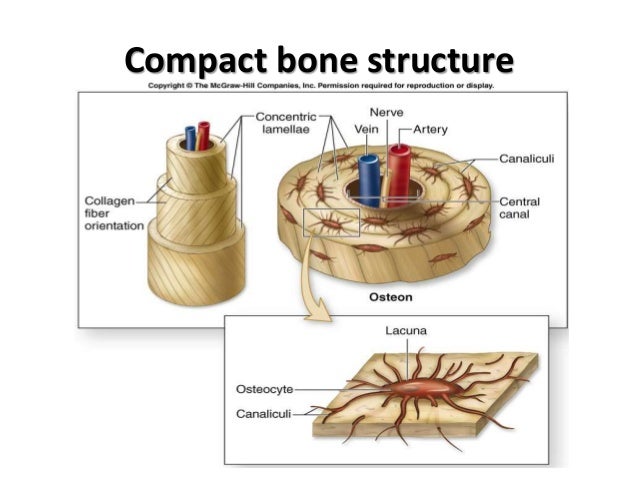
Cartilage + Inside of Bones - Anatomy & Physiology 508 ... from classconnection.s3.amazonaws.com The cells of compact bone, which is also called cortical bone, appear to be tightly packed into a solid mass. The two main structural components typically include spongy bone on the interior, with an outer layer of compact bone. Compact bone accounts for 80% of the bones in the human body. The diagram above shows a longitudinal view of an osteon. The periosteum then secretes compact bone superficial to the spongy bone. Haversian canals (sometimes canals of havers) are a series of microscopic tubes in the outermost region of bone called cortical bone. Structure of compact bone your skills & rank. Compact bone is formed from a number of osteons, which are circular units of bone material and blood vessels.
Bones are hard organs inside our bodies that make up our skeletal system.
They serve the purpose of protecting our bodies and also provide a structure and shape. It makes up the outer cortex of all bones and is in immediate contact with the periosteum. Start studying compact bone labeling. Compact bone is the strongest form of bone tissue containing few spaces. It can be found under the periosteum and in the diaphyses of long bones, where it provides support and protection. Some, mostly older, compact bone is remodelled to form these haversian systems (or osteons). The spongy bone crowds nearby blood vessels, which eventually condense into red bone marrow ( figure 6.4.1 d ). Human bone generally comprises osseous tissue, an outer coating called a periosteum, and bone marrow. Although the calls are close together, this type of bone is not completely solid. The key difference between compact bone and spongy bone is that the compact bone is a tough and heavy bone that forms the diaphysis of long bones while the spongy bone is a soft and light bone that forms the epiphysis of long bones. Related posts of compact bone diagram labeled anatomy of neck vein. The main function of compact bone is to support the whole body, whereas spongy bones support the body structure. The latter helps save materials, and provide movement to.
It can be found under the periosteum and in the diaphyses of long bones, where it provides support and protection. Compact bone, also called cortical bone, is the hard, stiff, smooth, thin, white bone tissue that surrounds all bones in the human body. There is a printable worksheet available for download here so you can take the quiz with pen and paper. The periosteum then secretes compact bone superficial to the spongy bone. (b) in this micrograph of the osteon, you can clearly see the concentric lamellae and central canals.
Structure Of Compact Bone And The Osteon - sharedoc from image.slidesharecdn.com Compact bone is the denser, stronger of the two types of osseous tissue (figure 6.3.6). Compact bone diagram osteon compact bone ap pinterest anatomy human anatomy and. Compact bone, also called cortical bone, dense bone in which the bony matrix is solidly filled with organic ground substance and inorganic salts, leaving only tiny spaces (lacunae) that contain the osteocytes, or bone cells.compact bone makes up 80 percent of the human skeleton; Structure of compact bone your skills & rank. Haversian canals (sometimes canals of havers) are a series of microscopic tubes in the outermost region of bone called cortical bone. Anatomy of neck vein 12 photos of the anatomy of neck vein anatomy internal jugular vein cannulation, anatomy of internal jugular vein in neck, anatomy of internal jugular vein pdf, anatomy of jugular vein in cattle, anatomy of the neck veins, human anatomy, anatomy internal jugular vein cannulation, anatomy of internal. The compact bone is composed of calcified extracellular material the bone matrix and 3 major cell types which are osteoblast which ssynthesize and secrete the organic components of bone matrix which include type 1 collagen fibers proteoglycans and several glycoproteins such as ostepnectin. The key difference between compact bone and spongy bone is that the compact bone is a tough and heavy bone that forms the diaphysis of long bones while the spongy bone is a soft and light bone that forms the epiphysis of long bones.
(b) in this micrograph of the osteon, you can clearly see the concentric lamellae and central canals.
Compact bone is formed from a number of osteons, which are circular units of bone material and blood vessels. (b) in this micrograph of the osteon, you can clearly see the concentric lamellae and central canals. Under periosteum of all bones is the bulk of the diaphysis of long bones. The diagram above shows a longitudinal view of an osteon. Provides protection and support while resisting stress from weight and movement. Compact bone is hard and forms the outer layer of any bone. It is also called osseous tissue or cortical bone and it provides structure and support for an organism as part of its skeleton, in addition to being a location for the storage of minerals like calcium.about 80% of the weight of the human skeleton comes from. Compact bone is the denser, stronger of the two types of osseous tissue (figure 6.3.6). The main function of compact bone is to support the whole body, whereas spongy bones support the body structure. Learn vocabulary, terms, and more with flashcards, games, and other study tools. A diagram of the anatomy of a bone, showing the compact bone. Structure of compact bone your skills & rank. About press copyright contact us creators advertise developers terms privacy policy & safety how youtube works test new features press copyright contact us creators.